ID:SATB1_HUMAN DESCRIPTION: RecName: Full=DNA-binding protein SATB1; AltName: Full=Special AT-rich sequence-binding protein 1; FUNCTION: Crucial silencing factor contributing to the initiation of X inactivation mediated by Xist RNA that occurs during embryogenesis and in lymphoma (By similarity). Binds to DNA at special AT-rich sequences, the consensus SATB1-binding sequence (CSBS), at nuclear matrix- or scaffold-associated regions. Thought to recognize the sugar-phosphate structure of double-stranded DNA. Transcriptional repressor controlling nuclear and viral gene expression in a phosphorylated and acetylated status-dependent manner, by binding to matrix attachment regions (MARs) of DNA and inducing a local chromatin-loop remodeling. Acts as a docking site for several chromatin remodeling enzymes (e.g. PML at the MHC-I locus) and also by recruiting corepressors (HDACs) or coactivators (HATs) directly to promoters and enhancers. Modulates genes that are essential in the maturation of the immune T-cell CD8SP from thymocytes. Required for the switching of fetal globin species, and beta- and gamma-globin genes regulation during erythroid differentiation. Plays a role in chromatin organization and nuclear architecture during apoptosis. Interacts with the unique region (UR) of cytomegalovirus (CMV). Alu-like motifs and SATB1- binding sites provide a unique chromatin context which seems preferentially targeted by the HIV-1 integration machinery. Moreover, HIV-1 Tat may overcome SATB1-mediated repression of IL2 and IL2RA (interleukin) in T-cells by binding to the same domain than HDAC1. Delineates specific epigenetic modifications at target gene loci, directly up-regulating metastasis-associated genes while down-regulating tumor-suppressor genes. Reprograms chromatin organization and the transcription profiles of breast tumors to promote growth and metastasis. SUBUNIT: Interacts (via DNA-binding domains) with CUX1; leading to inhibit the attachment to DNA (By similarity). Homodimer. Part of the nuclear protein complex gamma-globin promoter and enhancer binding factor (gamma-PE) composed at least by SATB1 and HOXB2. Interaction with CtBP1 when not acetylated stabalizes attachment to DNA and promotes transcription repression. Interacts with PCAF. Interacts with sumoylated PML, HDAC1 and HIV-1 Tat via the PDZ- like dimerization domain. Interacts also with DYNLT3 and POLR2J2. Binds to EP300. INTERACTION: P55212:CASP6; NbExp=2; IntAct=EBI-743747, EBI-718729; P63165:SUMO1; NbExp=2; IntAct=EBI-743747, EBI-80140; SUBCELLULAR LOCATION: Nucleus matrix. Nucleus, PML body. Note=Organized into a cage-like network anchoring loops of heterochromatin and tethering specialized DNA sequences. When sumoylated, localized in promyelocytic leukemia nuclear bodies (PML NBs). TISSUE SPECIFICITY: Expressed predominantly in thymus. PTM: Sumoylated. Sumoylation promotes cleavage by caspases. PTM: Phosphorylated by PKC. Acetylated by PCAF. Phosphorylated form interacts with HDAC1, but unphosphorylated form interacts with PCAF. DNA binding properties are activated by phosphorylation and inactivated by acetylation. In opposition, gene expression is down-regulated by phosphorylation but up-regulated by acetylation. PTM: Cleaved at Asp-254 by caspase-3 and caspase-6 during T-cell apoptosis in thymus and during B-cell stimulation. The cleaved forms can not dimerize and lose transcription regulation function because of impaired DNA and chromatin association. SIMILARITY: Belongs to the CUT homeobox family. SIMILARITY: Contains 2 CUT DNA-binding domains. SIMILARITY: Contains 1 homeobox DNA-binding domain. SEQUENCE CAUTION: Sequence=BAD92998.1; Type=Erroneous initiation;
The RNAfold program from the Vienna RNA Package is used to perform the secondary structure predictions and folding calculations. The estimated folding energy is in kcal/mol. The more negative the energy, the more secondary structure the RNA is likely to have.
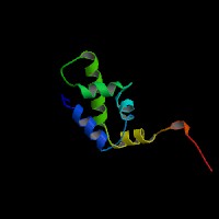
ModBase Predicted Comparative 3D Structure on Q01826
Front
Top
Side
The pictures above may be empty if there is no ModBase structure for the protein. The ModBase structure frequently covers just a fragment of the protein. You may be asked to log onto ModBase the first time you click on the pictures. It is simplest after logging in to just click on the picture again to get to the specific info on that model.
Orthologous Genes in Other Species
Orthologies between human, mouse, and rat are computed by taking the best BLASTP hit, and filtering out non-syntenic hits. For more distant species reciprocal-best BLASTP hits are used. Note that the absence of an ortholog in the table below may reflect incomplete annotations in the other species rather than a true absence of the orthologous gene.
Gene Ontology (GO) Annotations with Structured Vocabulary
Molecular Function: GO:0000977 RNA polymerase II regulatory region sequence-specific DNA binding GO:0000981 RNA polymerase II transcription factor activity, sequence-specific DNA binding GO:0001227 transcriptional repressor activity, RNA polymerase II transcription regulatory region sequence-specific binding GO:0003677 DNA binding GO:0003682 chromatin binding GO:0003690 double-stranded DNA binding GO:0003700 transcription factor activity, sequence-specific DNA binding GO:0005515 protein binding
Biological Process: GO:0000122 negative regulation of transcription from RNA polymerase II promoter GO:0006325 chromatin organization GO:0006338 chromatin remodeling GO:0006351 transcription, DNA-templated GO:0006355 regulation of transcription, DNA-templated GO:0008544 epidermis development GO:0016032 viral process GO:0016571 histone methylation GO:0042110 T cell activation GO:0043367 CD4-positive, alpha-beta T cell differentiation GO:0043374 CD8-positive, alpha-beta T cell differentiation GO:0050798 activated T cell proliferation GO:0060004 reflex
 Sequence and Links to Tools and Databases
  Common Gene Haplotype Alleles