ID:EPOR_HUMAN DESCRIPTION: RecName: Full=Erythropoietin receptor; Short=EPO-R; Flags: Precursor; FUNCTION: Receptor for erythropoietin. Mediates erythropoietin- induced erythroblast proliferation and differentiation. Upon EPO stimulation, EPOR dimerizes triggering the JAK2/STAT5 signaling cascade. In some cell types, can also activate STAT1 and STAT3. May also activate the LYN tyrosine kinase. FUNCTION: Isoform EPOR-T acts as a dominant-negative receptor of EPOR-mediated signaling. SUBUNIT: Forms homodimers on EPO stimulation. The tyrosine- phosphorylated form interacts with several SH2 domain-containing proteins including LYN (By similarity), the adapter protein APS, PTPN6 (By similarity), PTPN11, JAK2, PI3 kinases, STAT5A/B, SOCS3, CRKL (By similarity). Interacts with INPP5D/SHIP1 (By similarity). The N-terminal SH2 domain of PTPN6 binds Tyr-454 and inhibits signaling through dephosphorylation of JAK2 (By similarity). APS binding also inhibits the JAK-STAT signaling. Binding to PTPN11, preferentially through the N-terminal SH2 domain, promotes mitogenesis and phosphorylation of PTPN11 (By similarity). Binding of JAK2 (through its N-terminal) promotes cell-surface expression (By similarity). Interaction with the ubiquitin ligase NOSIP mediates EPO-induced cell proliferation. Interacts with ATXN2L. INTERACTION: Self; NbExp=2; IntAct=EBI-617321, EBI-617321; Q62225:Cish (xeno); NbExp=4; IntAct=EBI-617321, EBI-617489; P01588:EPO; NbExp=2; IntAct=EBI-617321, EBI-1027362; P16885:PLCG2; NbExp=3; IntAct=EBI-617321, EBI-617403; O14508:SOCS2; NbExp=3; IntAct=EBI-617321, EBI-617737; SUBCELLULAR LOCATION: Cell membrane; Single-pass type I membrane protein. SUBCELLULAR LOCATION: Isoform EPOR-S: Secreted. Note=Secreted and located to the cell surface. TISSUE SPECIFICITY: Erythroid cells and erythroid progenitor cells. Isoform EPOR-F is the most abundant form in EPO-dependent erythroleukemia cells and in late-stage erythroid progenitors. Isoform EPOR-S and isoform EPOR-T are the predominant forms in bone marrow. Isoform EPOR-T is the most abundant from in early- stage erythroid progenitor cells. DOMAIN: The WSXWS motif appears to be necessary for proper protein folding and thereby efficient intracellular transport and cell- surface receptor binding. DOMAIN: The box 1 motif is required for JAK interaction and/or activation. DOMAIN: Contains 1 copy of a cytoplasmic motif that is referred to as the immunoreceptor tyrosine-based inhibitor motif (ITIM). This motif is involved in modulation of cellular responses. The phosphorylated ITIM motif can bind the SH2 domain of several SH2- containing phosphatases. PTM: On EPO stimulation, phosphorylated on C-terminal tyrosine residues by JAK2. The phosphotyrosine motifs are also recruitment sites for several SH2-containing proteins and adapter proteins which mediate cell proliferation. Phosphorylation on Tyr-454 is required for PTPN6 interaction, Tyr-426 for PTPN11. Tyr-426 is also required for SOCS3 binding, but Tyr-454/Tyr-456 motif is the preferred binding site. PTM: Ubiquitination at Lys-281 mediates receptor internalization, whereas ubiquitination at Lys-453 promotes trafficking of activated receptors to the lysosomes for degradation (By similarity). Ubiquitinated by NOSIP; appears to be either multi- monoubiquitinated or polyubiquitinated. Ubiquitination mediates proliferation and survival of EPO-dependent cells. DISEASE: Defects in EPOR are the cause of familial erythrocytosis type 1 (ECYT1) [MIM:133100]. ECYT1 is an autosomal dominant disorder characterized by increased serum red blood cell mass, elevated hemoglobin and hematocrit, hypersensitivity of erythroid progenitors to erythropoietin, erythropoietin low serum levels, and no increase in platelets nor leukocytes. It has a relatively benign course and does not progress to leukemia. SIMILARITY: Belongs to the type I cytokine receptor family. Type 1 subfamily. SIMILARITY: Contains 1 fibronectin type-III domain.
The RNAfold program from the Vienna RNA Package is used to perform the secondary structure predictions and folding calculations. The estimated folding energy is in kcal/mol. The more negative the energy, the more secondary structure the RNA is likely to have.
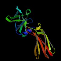
ModBase Predicted Comparative 3D Structure on P19235
Front
Top
Side
The pictures above may be empty if there is no ModBase structure for the protein. The ModBase structure frequently covers just a fragment of the protein. You may be asked to log onto ModBase the first time you click on the pictures. It is simplest after logging in to just click on the picture again to get to the specific info on that model.
Orthologous Genes in Other Species
Orthologies between human, mouse, and rat are computed by taking the best BLASTP hit, and filtering out non-syntenic hits. For more distant species reciprocal-best BLASTP hits are used. Note that the absence of an ortholog in the table below may reflect incomplete annotations in the other species rather than a true absence of the orthologous gene.
 Sequence and Links to Tools and Databases
  Common Gene Haplotype Alleles