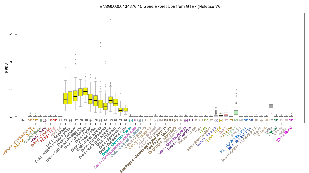
ID:CRUM1_HUMAN DESCRIPTION: RecName: Full=Protein crumbs homolog 1; Flags: Precursor; FUNCTION: Plays a role in photoreceptor morphogenesis in the retina. May maintain cell polarization and adhesion. SUBUNIT: Forms a complex with MPDZ (By similarity). Forms a complex with MPP4 and MPP5. SUBCELLULAR LOCATION: Isoform 1: Apical cell membrane; Single-pass type I membrane protein. Note=Distributed at the apical membrane of all retinal epithelial cells. Located in the apical membrane of the adherens junction in outer limiting membrane (OLM) of the retina. SUBCELLULAR LOCATION: Isoform 2: Secreted. TISSUE SPECIFICITY: Preferential expression in retina, also expressed in brain, testis, fetal brain and fetal eye. PTM: Extensively glycosylated. DISEASE: Note=CRB1 mutations have been found in various retinal dystrophies, chronic and disabling disorders of visual function. They predominantly involve the posterior portion of the ocular fundus, due to degeneration in the sensory layer of the retina, retinal pigment epithelium, Bruch membrane, choroid, or a combination of these tissues. Onset of inherited retinal dystrophies is painless, bilateral and typically progressive. Most people experience gradual peripheral vision loss or tunnel vision, and difficulties with poor illumination and night vision. Central vision is usually unaffected, so the person may still be able to read. However, it can also deteriorate to cause total blindness. Examples of retinal dystrophies are retinitis pigmentosa, Leber congenital amaurosis, cone-rod dystrophy among others. DISEASE: Defects in CRB1 are the cause of retinitis pigmentosa type 12 (RP12) [MIM:600105]. A retinal dystrophy belonging to the group of pigmentary retinopathies. Retinitis pigmentosa is characterized by retinal pigment deposits visible on fundus examination and primary loss of rod photoreceptor cells, followed by secondary loss of cone photoreceptors. Patients typically have night vision blindness and loss of midperipheral visual field. As their condition progresses, they lose their far peripheral visual field and eventually central vision as well. RP12 is an autosomal recessive severe form oFTen manifesting in early childhood. Patients experiment progressive visual field loss with severe visual impairment before the age of twenty. Some patients have a preserved paraarteriolar retinal pigment epithelium (PPRPE) and hypermetropia. DISEASE: Defects in CRB1 are the cause of Leber congenital amaurosis type 8 (LCA8) [MIM:613835]. LCA designates a clinically and genetically heterogeneous group of childhood retinal degenerations, generally inherited in an autosomal recessive manner. Affected infants have little or no retinal photoreceptor function as tested by electroretinography. LCA represents the most common genetic cause of congenital visual impairment in infants and children. DISEASE: Defects in CRB1 are the cause of pigmented paravenous chorioretinal atrophy (PPCRA) [MIM:172870]. PPCRA is an unusual retinal degeneration characterized by accumulation of pigmentation along retinal veins. PPCRA is dominantly inherited, but exhibited variable expressivity. Males are more likely to exhibit a severe phenotype, whereas females may remain virtually asymptomatic even in later years. The PPCRA phenotype is associated with a mutation in CRB1 gene which is likely to affect the structure of the CRB1 protein. SIMILARITY: Belongs to the Crumbs protein family. SIMILARITY: Contains 19 EGF-like domains. SIMILARITY: Contains 3 laminin G-like domains. SEQUENCE CAUTION: Sequence=CAE45845.1; Type=Erroneous termination; Positions=567; Note=Translated as Trp; Sequence=CAI16644.1; Type=Erroneous gene model prediction; WEB RESOURCE: Name=Mutations of the CRB1 gene; Note=Retina International's Scientific Newsletter; URL="http://www.retina-international.org/files/sci-news/crb1mut.htm"; WEB RESOURCE: Name=GeneReviews; URL="http://www.ncbi.nlm.nih.gov/sites/GeneTests/lab/gene/CRB1";
The RNAfold program from the Vienna RNA Package is used to perform the secondary structure predictions and folding calculations. The estimated folding energy is in kcal/mol. The more negative the energy, the more secondary structure the RNA is likely to have.
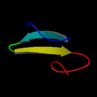
ModBase Predicted Comparative 3D Structure on P82279
Front
Top
Side
The pictures above may be empty if there is no ModBase structure for the protein. The ModBase structure frequently covers just a fragment of the protein. You may be asked to log onto ModBase the first time you click on the pictures. It is simplest after logging in to just click on the picture again to get to the specific info on that model.
Orthologous Genes in Other Species
Orthologies between human, mouse, and rat are computed by taking the best BLASTP hit, and filtering out non-syntenic hits. For more distant species reciprocal-best BLASTP hits are used. Note that the absence of an ortholog in the table below may reflect incomplete annotations in the other species rather than a true absence of the orthologous gene.
 Sequence and Links to Tools and Databases
  Common Gene Haplotype Alleles