ID:CFAH_HUMAN DESCRIPTION: RecName: Full=Complement factor H; AltName: Full=H factor 1; Flags: Precursor; FUNCTION: Factor H functions as a cofactor in the inactivation of C3b by factor I and also increases the rate of dissociation of the C3bBb complex (C3 convertase) and the (C3b)NBB complex (C5 convertase) in the alternative complement pathway. SUBCELLULAR LOCATION: Secreted. TISSUE SPECIFICITY: Expressed by the liver and secreted in plasma. DISEASE: Genetic variations in CFH are associated with basal laminar drusen (BLD) [MIM:126700]; also known as drusen of Bruch membrane or cuticular drusen or grouped early adult-onset drusen. Drusen are extracellular deposits that accumulate below the retinal pigment epithelium on Bruch membrane. Basal laminar drusen refers to an early adult-onset drusen phenotype that shows a pattern of uniform small, slightly raised yellow subretinal nodules randomly scattered in the macula. In later stages, these drusen often become more numerous, with clustered groups of drusen scattered throughout the retina. In time these small basal laminar drusen may expand and ultimately lead to a serous pigment epithelial detachment of the macula that may result in vision loss. DISEASE: Defects in CFH are the cause of complement factor H deficiency (CFHD) [MIM:609814]. A disorder that can manifest as several different phenotypes, including asymptomatic, recurrent bacterial infections, and renal failure. Laboratory features usually include decreased serum levels of factor H, complement component C3, and a decrease in other terminal complement components, indicating activation of the alternative complement pathway. It is associated with a number of renal diseases with variable clinical presentation and progression, including membranoproliferative glomerulonephritis and atypical hemolytic uremic syndrome. DISEASE: Defects in CFH are a cause of susceptibility to hemolytic uremic syndrome atypical type 1 (AHUS1) [MIM:235400]. An atypical form of hemolytic uremic syndrome. It is a complex genetic disease characterized by microangiopathic hemolytic anemia, thrombocytopenia, renal failure and absence of episodes of enterocolitis and diarrhea. In contrast to typical hemolytic uremic syndrome, atypical forms have a poorer prognosis, with higher death rates and frequent progression to end-stage renal disease. Note=Susceptibility to the development of atypical hemolytic uremic syndrome can be conferred by mutations in various components of or regulatory factors in the complement cascade system. Other genes may play a role in modifying the phenotype. DISEASE: Genetic variation in CFH is associated with age-related macular degeneration type 4 (ARMD4) [MIM:610698]. ARMD is a multifactorial eye disease and the most common cause of irreversible vision loss in the developed world. In most patients, the disease is manifest as ophthalmoscopically visible yellowish accumulations of protein and lipid (known as drusen) that lie beneath the retinal pigment epithelium and within an elastin- containing structure known as Bruch membrane. SIMILARITY: Contains 20 Sushi (CCP/SCR) domains. SEQUENCE CAUTION: Sequence=CAB41739.1; Type=Frameshift; Positions=341; WEB RESOURCE: Name=CFHbase; Note=CFH mutation db; URL="http://bioinf.uta.fi/CFHbase/"; WEB RESOURCE: Name=GeneReviews; URL="http://www.ncbi.nlm.nih.gov/sites/GeneTests/lab/gene/CFH"; WEB RESOURCE: Name=SeattleSNPs; URL="http://pga.gs.washington.edu/data/cfh/";
The RNAfold program from the Vienna RNA Package is used to perform the secondary structure predictions and folding calculations. The estimated folding energy is in kcal/mol. The more negative the energy, the more secondary structure the RNA is likely to have.
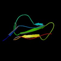
ModBase Predicted Comparative 3D Structure on P08603
Front
Top
Side
The pictures above may be empty if there is no ModBase structure for the protein. The ModBase structure frequently covers just a fragment of the protein. You may be asked to log onto ModBase the first time you click on the pictures. It is simplest after logging in to just click on the picture again to get to the specific info on that model.
Orthologous Genes in Other Species
Orthologies between human, mouse, and rat are computed by taking the best BLASTP hit, and filtering out non-syntenic hits. For more distant species reciprocal-best BLASTP hits are used. Note that the absence of an ortholog in the table below may reflect incomplete annotations in the other species rather than a true absence of the orthologous gene.
 Sequence and Links to Tools and Databases
  Common Gene Haplotype Alleles