ID:CD209_HUMAN DESCRIPTION: RecName: Full=CD209 antigen; AltName: Full=C-type lectin domain family 4 member L; AltName: Full=Dendritic cell-specific ICAM-3-grabbing non-integrin 1; Short=DC-SIGN; Short=DC-SIGN1; AltName: CD_antigen=CD209; FUNCTION: Pathogen-recognition receptor expressed on the surface of immature dendritic cells (DCs) and involved in initiation of primary immune response. Thought to mediate the endocytosis of pathogens which are subsequently degraded in lysosomal compartments. The receptor returns to the cell membrane surface and the pathogen-derived antigens are presented to resting T-cells via MHC class II proteins to initiate the adaptive immune response. Probably recognizes in a calcium-dependent manner high mannose N-linked oligosaccharides in a variety of pathogen antigens, including HIV-1 gp120, HIV-2 gp120, SIV gp120, ebolavirus glycoproteins, cytomegalovirus gB, HCV E2, dengue virus gE, Leishmania pifanoi LPG, Lewis-x antigen in Helicobacter pylori LPS, mannose in Klebsiella pneumonae LPS, di-mannose and tri- mannose in Mycobacterium tuberculosis ManLAM and Lewis-x antigen in Schistosoma mansoni SEA. FUNCTION: On DCs it is a high affinity receptor for ICAM2 and ICAM3 by binding to mannose-like carbohydrates. May act as a DC rolling receptor that mediates transendothelial migration of DC presursors from blood to tissues by binding endothelial ICAM2. Seems to regulate DC-induced T-cell proliferation by binding to ICAM3 on T-cells in the immunological synapse formed between DC and T-cells. SUBUNIT: Homotetramer. Binds to many viral surface glycoproteins such as HIV-1 gp120, HIV-2 gp120, SIV gp120, ebolavirus envelope glycoproteins, cytomegalovirus gB, HCV E2 and dengue virus major envelope protein E. SUBCELLULAR LOCATION: Isoform 1: Cell membrane; Single-pass type II membrane protein (Probable). SUBCELLULAR LOCATION: Isoform 2: Cell membrane; Single-pass type II membrane protein (Probable). SUBCELLULAR LOCATION: Isoform 3: Cell membrane; Single-pass type II membrane protein (Probable). SUBCELLULAR LOCATION: Isoform 4: Cell membrane; Single-pass type II membrane protein (Probable). SUBCELLULAR LOCATION: Isoform 5: Cell membrane; Single-pass type II membrane protein (Probable). SUBCELLULAR LOCATION: Isoform 6: Secreted (Probable). SUBCELLULAR LOCATION: Isoform 7: Secreted (Probable). SUBCELLULAR LOCATION: Isoform 8: Secreted (Probable). SUBCELLULAR LOCATION: Isoform 9: Secreted (Probable). SUBCELLULAR LOCATION: Isoform 10: Secreted (Probable). SUBCELLULAR LOCATION: Isoform 11: Secreted (Probable). SUBCELLULAR LOCATION: Isoform 12: Secreted (Probable). TISSUE SPECIFICITY: Predominantly expressed in dendritic cells and in DC-residing tissues. Also found in placental macrophages, endothelial cells of placental vascular channels, peripheral blood mononuclear cells, and THP-1 monocytes. DOMAIN: The tandem repeat domain, also called neck domain, mediates oligomerization. POLYMORPHISM: Genetic variations in CD209 determine Mycobacterium tuberculosis susceptibility [MIM:607948]. POLYMORPHISM: Genetic variations in CD209 may influence susceptibility or resistance to dengue virus infection, as well as disease progression and severity [MIM:614371]. A promoter polymorphism in the CD209 gene is associated with protection from dengue fever, but not dengue hemorrhagic fever. MISCELLANEOUS: In vitro, is a receptor for HIV-1 and transmits HIV-1 either in trans without DC infection, or in cis following a DC infection to permissive T-cells to induce a robust infection. Bound HIV-1 remains infectious over a prolonged period of time and it is proposed that bound HIV-1 is not degraded but protected in non-lysosomal acidic organelles within the DCs close to the cell membrane thus contributing to the HIV-1 infectious potential during transport by DCs from the periphery to lymphoid organs. SIMILARITY: Contains 1 C-type lectin domain. SEQUENCE CAUTION: Sequence=AAK91858.1; Type=Miscellaneous discrepancy; Note=Aberrant splicing; WEB RESOURCE: Name=Wikipedia; Note=DC-SIGN entry; URL="http://en.wikipedia.org/wiki/DC-SIGN"; WEB RESOURCE: Name=Functional Glycomics Gateway - Glycan Binding; Note=DC-SIGN; URL="http://www.functionalglycomics.org/glycomics/GBPServlet?&operationType=view&cbpId=cbp_hum_Ctlect_00121";
The RNAfold program from the Vienna RNA Package is used to perform the secondary structure predictions and folding calculations. The estimated folding energy is in kcal/mol. The more negative the energy, the more secondary structure the RNA is likely to have.
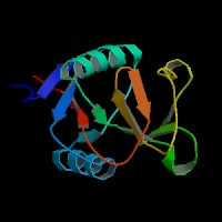
ModBase Predicted Comparative 3D Structure on Q9NNX6
Front
Top
Side
The pictures above may be empty if there is no ModBase structure for the protein. The ModBase structure frequently covers just a fragment of the protein. You may be asked to log onto ModBase the first time you click on the pictures. It is simplest after logging in to just click on the picture again to get to the specific info on that model.
Orthologous Genes in Other Species
Orthologies between human, mouse, and rat are computed by taking the best BLASTP hit, and filtering out non-syntenic hits. For more distant species reciprocal-best BLASTP hits are used. Note that the absence of an ortholog in the table below may reflect incomplete annotations in the other species rather than a true absence of the orthologous gene.
 Sequence and Links to Tools and Databases
  Common Gene Haplotype Alleles