ID:PI42A_HUMAN DESCRIPTION: RecName: Full=Phosphatidylinositol 5-phosphate 4-kinase type-2 alpha; EC=2.7.1.149; AltName: Full=1-phosphatidylinositol 5-phosphate 4-kinase 2-alpha; AltName: Full=Diphosphoinositide kinase 2-alpha; AltName: Full=PIP5KIII; AltName: Full=Phosphatidylinositol 5-phosphate 4-kinase type II alpha; Short=PI(5)P 4-kinase type II alpha; Short=PIP4KII-alpha; AltName: Full=PtdIns(4)P-5-kinase B isoform; AltName: Full=PtdIns(4)P-5-kinase C isoform; AltName: Full=PtdIns(5)P-4-kinase isoform 2-alpha; FUNCTION: Catalyzes the phosphorylation of phosphatidylinositol 5- phosphate (PtdIns5P) on the fourth hydroxyl of the myo-inositol ring, to form phosphatidylinositol 4,5-bisphosphate (PtdIns(4,5)P2). May exert its function by regulating the levels of PtdIns5P, which functions in the cytosol by increasing AKT activity and in the nucleus signals through ING2. May regulate the pool of cytosolic PtdIns5P in response to the activation of tyrosine phosphorylation. May negatively regulate insulin- stimulated glucose uptake by lowering the levels of PtdIns5P. May be involved in thrombopoiesis, and the terminal maturation of megakaryocytes and regulation of their size. CATALYTIC ACTIVITY: ATP + 1-phosphatidyl-1D-myo-inositol 5- phosphate = ADP + 1-phosphatidyl-1D-myo-inositol 4,5-bisphosphate. BIOPHYSICOCHEMICAL PROPERTIES: Kinetic parameters: KM=50 uM for phosphatidylinositol-5- phosphate; Vmax=466 pmol/min/ug enzyme; SUBUNIT: Homodimer (By similarity). Interacts with PIP4K2B. Interaction with PIP4K2B may regulate localization to the nucleus. SUBCELLULAR LOCATION: Cell membrane (By similarity). Nucleus. Cytoplasm. Note=May translocate from the cytosol to the cell membrane upon activation of tyrosine phosphorylation. May translocate from the inner to the outer segments of the rod photoreceptor cells in response to light (By similarity). Localization to the nucleus is modulated by the interaction with PIP4K2B. TISSUE SPECIFICITY: Expressed ubiquitously, with high levels in the brain. Present in most tissues, except notably skeletal muscle and small intestine. SIMILARITY: Contains 1 PIPK domain. CAUTION: This protein was previously thought to be a phosphatidylinositol 4-phosphate 5-kinase.
The RNAfold program from the Vienna RNA Package is used to perform the secondary structure predictions and folding calculations. The estimated folding energy is in kcal/mol. The more negative the energy, the more secondary structure the RNA is likely to have.
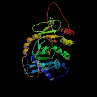
ModBase Predicted Comparative 3D Structure on P48426
Front
Top
Side
The pictures above may be empty if there is no ModBase structure for the protein. The ModBase structure frequently covers just a fragment of the protein. You may be asked to log onto ModBase the first time you click on the pictures. It is simplest after logging in to just click on the picture again to get to the specific info on that model.
Orthologous Genes in Other Species
Orthologies between human, mouse, and rat are computed by taking the best BLASTP hit, and filtering out non-syntenic hits. For more distant species reciprocal-best BLASTP hits are used. Note that the absence of an ortholog in the table below may reflect incomplete annotations in the other species rather than a true absence of the orthologous gene.
 Sequence and Links to Tools and Databases
  Common Gene Haplotype Alleles