ID:LCAT_HUMAN DESCRIPTION: RecName: Full=Phosphatidylcholine-sterol acyltransferase; EC=2.3.1.43; AltName: Full=Lecithin-cholesterol acyltransferase; AltName: Full=Phospholipid-cholesterol acyltransferase; Flags: Precursor; FUNCTION: Central enzyme in the extracellular metabolism of plasma lipoproteins. Synthesized mainly in the liver and secreted into plasma where it converts cholesterol and phosphatidylcholines (lecithins) to cholesteryl esters and lysophosphatidylcholines on the surface of high and low density lipoproteins (HDLs and LDLs). The cholesterol ester is then transported back to the liver. Has a preference for plasma 16:0-18:2 or 18:O-18:2 phosphatidylcholines. Also produced in the brain by primary astrocytes, and esterifies free cholesterol on nascent APOE-containing lipoproteins secreted from glia and influences cerebral spinal fluid (CSF) APOE- and APOA1 levels. Together with APOE and the cholesterol transporter ABCA1, plays a key role in the maturation of glial-derived, nascent lipoproteins. Required for remodeling high-density lipoprotein particles into their spherical forms. CATALYTIC ACTIVITY: Phosphatidylcholine + a sterol = 1- acylglycerophosphocholine + a sterol ester. ENZYME REGULATION: APOA1 is the most potent activator in plasma. Also activated by APOE, APOC1 and APOA4. BIOPHYSICOCHEMICAL PROPERTIES: Kinetic parameters: KM=0.97 mM for LDL; KM=0.4 mM for HDL(2); KM=0.10 mM for HDL(3); Vmax=8.3 mmol/min/mg enzyme with LDL as substrate; Vmax=0.58 mmol/min/mg enzyme with HDL(2) as substrate; Vmax=2.0 mmol/min/mg enzyme with HDL(3) as substrate; Note=Affinity for LDL is 2.3 to 4-fold lower than for HDL. Relative reactivities are 16% for HDL(3), 1.3% for HDL(2) and 6.5% for LDL; SUBCELLULAR LOCATION: Secreted. Note=Secreted into blood plasma. Produced in astrocytes and secreted into cerebral spinal fluid (CSF). TISSUE SPECIFICITY: Expressed mainly in brain, liver and testes. Secreted into plasma and cerebral spinal fluid. Expressed in Hep- G2 cell line. PTM: O- and N-glycosylated. O-glycosylation on Thr-431 and Ser-433 consists of sialylated galactose beta 1-->3N-acetylgalactosamine structures. N-glycosylated sites contain sialylated triantennary and/or biantennary complex structures. DISEASE: Defects in LCAT are the cause of lecithin-cholesterol acyltransferase deficiency (LCATD) [MIM:245900]; also called Norum disease. LCATD is a disorder of lipoprotein metabolism characterized by inadequate esterification of plasmatic cholesterol. Two clinical forms are recognized: familial LCAT deficiency and fish-eye disease. Familial LCAT deficiency is associated with a complete absence of alpha and beta LCAT activities and results in esterification anomalies involving both HDL (alpha-LCAT activity) and LDL (beta-LCAT activity). It causes a typical triad of diffuse corneal opacities, target cell hemolytic anemia, and proteinuria with renal failure. DISEASE: Defects in LCAT are a cause of fish-eye disease (FED) [MIM:136120]; also known as dyslipoproteinemic corneal dystrophy or alpha-LCAT deficiency. FED is due to a partial LCAT deficiency that affects only alpha-LCAT activity. It is characterized by low plasma HDL and corneal opacities due to accumulation of cholesterol deposits in the cornea ('fish-eye'). MISCELLANEOUS: Levels of LCAT activity correlates inversely with leptin levels as well as with obesity for a wide range of BMI values. SIMILARITY: Belongs to the AB hydrolase superfamily. Lipase family. WEB RESOURCE: Name=GeneReviews; URL="http://www.ncbi.nlm.nih.gov/sites/GeneTests/lab/gene/LCAT"; WEB RESOURCE: Name=Wikipedia; Note=Lecithin-cholesterol acyltransferase entry; URL="http://en.wikipedia.org/wiki/Lecithin-cholesterol_acyltransferase";
The RNAfold program from the Vienna RNA Package is used to perform the secondary structure predictions and folding calculations. The estimated folding energy is in kcal/mol. The more negative the energy, the more secondary structure the RNA is likely to have.
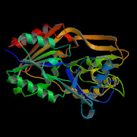
ModBase Predicted Comparative 3D Structure on P04180
Front
Top
Side
The pictures above may be empty if there is no ModBase structure for the protein. The ModBase structure frequently covers just a fragment of the protein. You may be asked to log onto ModBase the first time you click on the pictures. It is simplest after logging in to just click on the picture again to get to the specific info on that model.
Orthologous Genes in Other Species
Orthologies between human, mouse, and rat are computed by taking the best BLASTP hit, and filtering out non-syntenic hits. For more distant species reciprocal-best BLASTP hits are used. Note that the absence of an ortholog in the table below may reflect incomplete annotations in the other species rather than a true absence of the orthologous gene.
 Sequence and Links to Tools and Databases
  Common Gene Haplotype Alleles